SpecialCases
Right Submandiular Gland Sialography
Patient complains of swelling in relation to right lower back region of floor of mouth for last 1 month. Intraoral examination revealed healthy teeth and periodontium. Right submandibular gland region was tender on palpation.

Dilatation of right submandibular duct using lacrimal probe.

Canulation of right submandibular duct for injecting contrast media in the gland.

Radiograph revealing normal “Bush in winter” architecture of right submandibular gland.
Scientific Presentation by Post Graduate Students in Interdepartmental Meeting
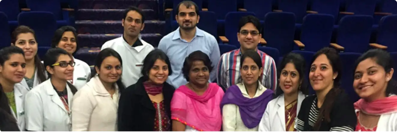
Dr Adrita Nag (2nd year PG) and Dr Ankur Bhagat (2nd year PG) presented a scientific presentation titled“Facial dystonia’s simplified! A common approach to an uncommon disease” in interdepartmental meeting held in Medical College Auditorium, SGT University on 26th February, 2016.This highly informative scientific presentation was attended by all the respected faculty members and post graduate students of Faculty of Dental Sciences, SGT University. It was a great learning experience involving a multidisciplinary management approach for optimum patient care.
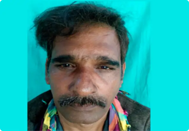
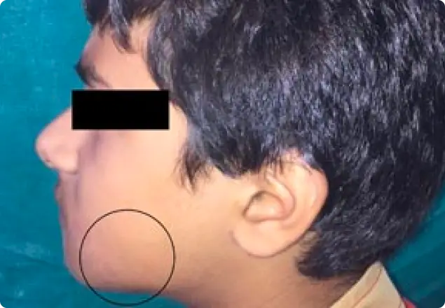
Hemi- Facial Microsomia
Patient reported to the department with complaint of difficulty in eating food due to irregularly arranged teeth.
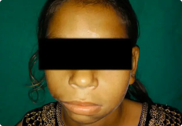
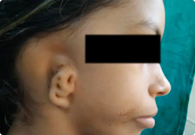
Extra-oral examination revealed prominent forehead, receded hairline, right ear smaller in size (microtia), malformed with a roughly oval shape and 3-4 blind fistulas overlying the ear (atresia). A horizontal scar mark measuring 18-20 mm in size seen extending from the right angle of the mouth till a line drawn perpendicular to the lateral canthus of the eye suggestive of lateral facial cleft. Right side mandible smaller in size, head of the right condyle is not palpable, hypotrophy appreciated in right masseter muscle. Intra oral examination revealed severe malocclusion with interdental spacing between anterior maxillary teeth suggestive of developing malocclusion.
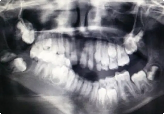
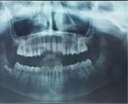
Panoramic radiograph reveals mixed dentition phase with multiple developing tooth buds. Zygomatic process cannot be traced completely on the right side. The right half of the mandible from the symphysis region appears sharply bent upto the angle region. Right side temporo-mandibular joint is not appreciable,
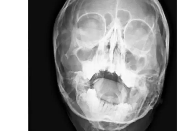
PA skull radiograph revealed an asymmetry on the right side with hypoplastic ramus, hypoplastic condylar head and aplastic coronoid process. The similar findings were confirmed by CT scan. Final diagnosis: Hemifacial Microsomia on right side OMENS Classification: O0M2BE3N71S2Management Immediate treatment: Orthodontic Opinion Planned treatment: Mandibular distraction osteogenesis.
Arterio-venous malformation involving left lower back maxillofacial region.
Patient reported to the department with complaint of difficulty in eating food due to irregularly arranged teeth.
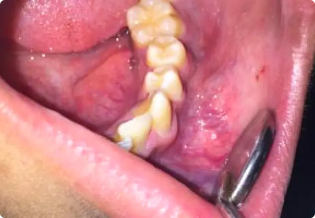
A bluish red bulbous swelling seen in the lower left buccal vestibule measuring about 3 x 2 cm with engorged capillaries along the periphery of lesion superiorly. Swelling was firm, compressible and with marked pulsations. Vestibular obliteration was present
Panoramic radiograph revealing a well defined radiolucency in the left mandibular body and anterior ramus region, causing root resorption of the involved teeth.
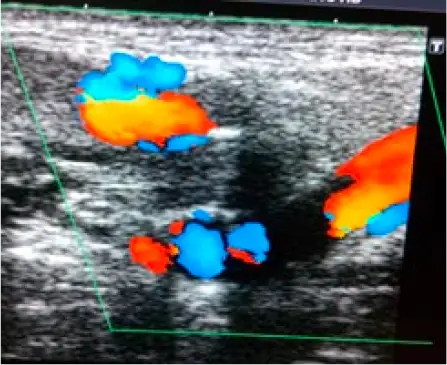
Colour doppler of left submandibular region revealing prominent vessels and biphasic, triphasic and venous pattern on spectral doppler, suggestive of Arterio-venous malformation.
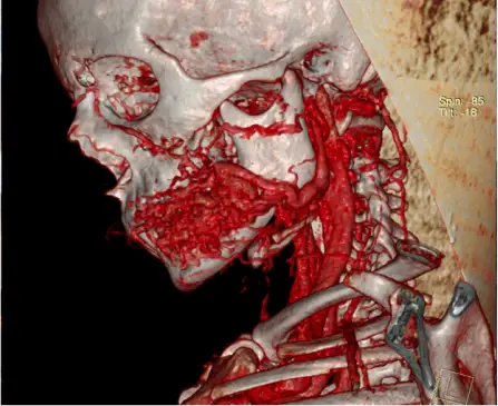
CT angiography confirming Arterio-venous malformation involving left lower back maxillofacial region.
Conclusion
The case highlights the various clinical and radiographic manifestations of arterio-venous malformations of maxillofacial region. Such cases should be accurately diagnosed and meticulously managed with a multi-disciplinary approach.
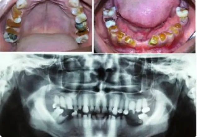
Dentinogenesis Imperfecta
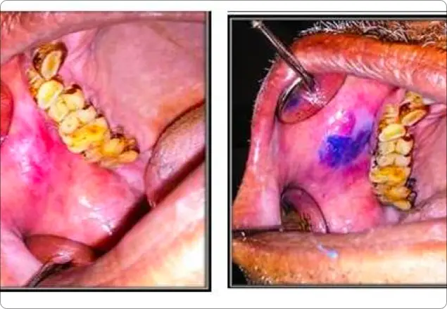
Toludine Blue Staining
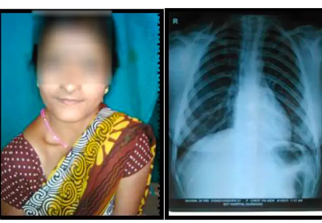
Cleidocranial Dysplasia
AV Malformation of Left Side of Face